Human-mouse chimera, the secrets of Tutankhamun's tomb, the fight against cancer
Interesting science news of the week
Human brain cells are transplanted into a mouse's brain, and the resulting chimera is used to study drugs for neurodegenerative diseases. From stem cells obtained from umbilical cord blood, an immune preparation was created to fight various types of cancer. Behind the wall of Tutankhamun's tomb, there may be hidden chambers, in one of which Nefertiti may be buried. Antisense oligonucleotide therapy for the first time helped a person with a disease like Stephen Hawking's. Scientists have created a neuroimplant that displays on a screen, with the help of an avatar, the words and gestures of completely paralyzed people.
Human cells in a mouse's brain — a medical breakthrough or an ethical problem?
This week an article was published about the largest-scale integration to date of human brain cells into an animal's body. This was done by a group of American scientists who study neurodegenerative diseases in a laboratory on the basis of grown human brain tissues.
Animals created in this way represent a unique material for testing preparations and for studying diseases connected with abnormal brain development, such as cerebral palsy. However, a number of researchers raise the ethical question of creating such chimeras (essentially, hybrids of a human and a mouse). Incidentally, all the necessary commissions approved the described experiment.
The human brain organoids that were implanted into the mouse pups represent clusters of neurons grown from human stem cells in Petri dishes. The transplantation was carried out a few days after the birth of the mice, when the main neural connections in the brain had already been formed. And this guaranteed that the human cells could not take on complex thought processes. Behavioral tests showed that they did not improve the intelligence of the rodents.
That is, the goal of creating the human-mouse chimera was not to create a supermouse, but to place tissues of the human brain in a realistic environment of a living organism. After all, neurobiologists suffered for decades from the impossibility of studying living tissue of the human brain. Organoids grown in Petri dishes have no body, cannot receive or send signals, and have no blood vessels.

The problem was conceived to be solved back in 2022 by transplanting brain organoids into newborn rats. The neurons matured and formed sensory pathways. And a couple of years later these rats were used to evaluate preparations against one of the severe genetic diseases. But then scientists had to squeeze the human transplant next to the rodent's own brain tissue, which grew quickly and occupied most of the available space.
In the new experiment the mice were genetically modified in advance: the precursor cells from which the cerebral cortex was supposed to form did not survive. And that means part of the brain cavity remained empty. In the absence of competition for space, the human cells grew over just three months, the tissue increased in volume fivefold and sent processes deep into the rodents' spinal cord. From the organoids full-fledged neurons developed, including specialized neurons of exactly the type that are most susceptible to neurodegenerative diseases. For example, to dementia.
Several experiments showed that the transplant can imitate a human disease: for this, for example, the mice were subjected to hypoxia — and their coordination of movements was impaired. Just like in people with cerebral palsy obtained as a result of prematurity. And since the transplant can imitate a disease — that means treatment methods can also be tested on it. Neurobiologists around the world are watching these experiments and weighing the ethical possibilities. After all, it is one thing — mice. And what will happen if it comes to primates?
T-cells created from umbilical cord blood were taught to recognize cancer in two ways
Scientists from the University of California in Los Angeles have developed T-cells that can recognize and attack solid tumors. The new method is designed for creating a ready-made cell preparation that can potentially be produced in advance and stored until the moment of use.
One of the main difficulties of modern cellular immunotherapy lies in the fact that the preparation has to be obtained individually for each patient. The use of mature T-lymphocytes from healthy donors is also limited: they can be attacked by the recipient's immune system, or they can attack healthy organ systems. The American scientists solved the problem by working on the immune cells at an earlier stage of their development. They genetically modified the stem cells of umbilical cord blood before those turned into mature T-lymphocytes. A gene of a receptor capable of recognizing the protein NY-ESO-1 was built into the cells. This molecule is found in the cells of many types of cancer — for example, melanoma, ovarian and prostate cancer.
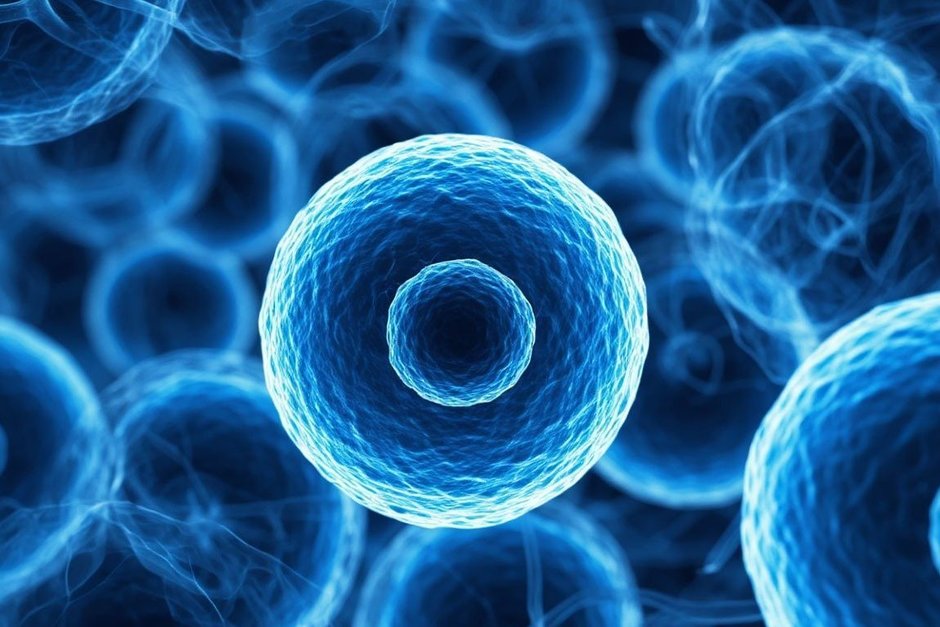
But what if the tumor “hides” from the T-cells trained on it by shedding that very marker protein? To prevent this, the researchers provided for a backup mechanism for detecting the tumor. In the modified T-cells they preserved the properties of natural killers — a reaction to stress signals characteristic of cancer cells. Such a combination should help fight the tumor even if it stops keeping on its surface the protein that the main receptor is oriented toward.
The effect of the new therapy was checked on mice. After a single injection the introduced immune cells began to multiply well — their number quickly grew a hundredfold. They held back the growth of tumors for a long time, and the experimental animals lived longer. At the same time the researchers did not record dangerous side effects.
Another advantage of the technology is connected with scaling up production. According to the authors' estimate, a small amount of umbilical cord blood stem cells is enough to obtain trillions of immune cells in about six weeks. This, as the scientists believe, will be enough for manufacturing thousands of doses of the preparation with an approximate cost of about $5,000 each. Currently the technology is being prepared for clinical trials on humans.
New secrets of Tutankhamun's tomb
The tomb of Tutankhamun in Egypt's Valley of the Kings will not stop stirring the souls of archaeologists. Many of them believe that behind its walls there are hidden chambers, in which, presumably, Nefertiti is buried. Until now this remains only unproven suppositions. And now another study has come out, in which researchers claim that they have found new arguments in favor of this dubious but so tempting theory.
A group of scientists from Ain Shams University in Cairo conducted geophysical studies, including for the first time using gravitational methods to determine the density of the soil around the excavation site, and also applied ground-penetrating radar from inside the tomb. The newly minted Indiana Joneses claim that their data, in combination with studies of the structure of the tomb, the soil around it and the wall painting, convincingly testify to the presence of a hidden complex of corridors and rooms that were created even before the burial of Tutankhamun. The researchers are sure that the ground-penetrating radar shows a two-meter corridor leading from the burial chamber, which is densely filled with debris. And microgravitational studies show the presence of structures that can be convincingly qualified as chambers filled with some kind of content.

This study became the next in a series of attempts to imagine what is hidden behind the walls of Tutankhamun's tomb. Let us recall that it was found in 1922, and it was built in the 14th century BC. Many archaeologists believe that for a royal burial it is too modest in size, and on its northern wall there are suspicious lines and cracks. Back in 2015 the well-known British Egyptologist Nicholas Reeves suggested that this is a false wall, behind which the burial of the famous Nefertiti, the predecessor of Tutankhamun, may be hidden (many Egyptologists believe that she ruled Egypt as pharaoh for some time). Reeves believes that initially this tomb was large, but not designed for Tutankhamun. According to his version, after the unexpected death of the young pharaoh there was no ready tomb for him, and it was decided simply to expand the entrance of an already used one, arrange a burial in it, and block the deeper sections.
The decision on whether to look for these chambers a few meters from the existing tomb is in the hands of the Supreme Council of Antiquities of Egypt. If it is positive, careful drilling and the deployment of miniature tractors equipped with cameras may begin as early as November. The journal Nature wrote about this this week, citing the researchers who put forward new evidence.
Antisense oligonucleotide therapy for the first time helped with ALS
Scientists for the first time created an antisense preparation specially for one patient with a rare form of amyotrophic lateral sclerosis (ALS). After a year of treatment his condition improved: he continues to work as a doctor, and the indicators connected with damage to nerve cells returned to the normal range.
ALS is a severe disease of the nervous system, in which motor neurons gradually die — the nerve cells that control muscles. The most famous patient with this disease is Stephen Hawking. In the case described in this article, the cause of the disease was a rare mutation in the CHCHD10 gene. Because of it, a pathological protein accumulates in the cells, which kills motor neurons. In the end the person dies because of respiratory failure: he does not have enough strength even to inhale.
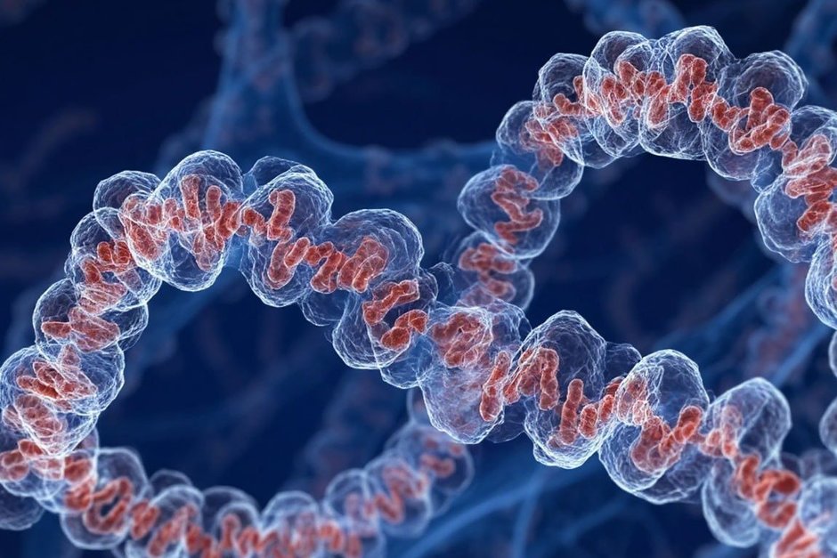
The researchers decided to act directly on this genetic mechanism. For the patient a preparation was created on the basis of an antisense oligonucleotide (ASO) — a short artificial molecule similar to a fragment of genetic material.
To understand the principle of its work, it is enough to imagine a gene as an instruction for producing a protein. First a copy is made from this instruction — messenger RNA, or mRNA. Then the cell uses it as a kind of note for producing the protein. The ASO finds the needed mRNA and binds to it. As a result the cell destroys this molecule and produces less pathological protein.
The scientists tested 320 variants of ASO and chose the most suitable in terms of specificity and safety. Then the patient received six injections of the preparation directly into the cerebrospinal fluid over the course of a year: first three doses of 50 mg, then three of 75 mg.
The treatment was tolerated well. After 12 months the concentration of the protein that is released upon damage to nerve cells and is used as an indicator of ALS activity — decreased by about 50% and entered the normal range. The clinical indicators also changed. Tests evaluating motor function, breathing and neurological state showed stability or moderate improvement. Cognitive functions and breathing also remain stable. The man continued to work as a doctor.
Especially noteworthy is that from the establishment of the genetic diagnosis to the creation of the preparation about three years passed. For comparison, the development of antisense preparations against more common mutations causing ALS previously took no less than ten years.
The researchers emphasize that for now it is a question of only one patient, so it is premature to draw conclusions about the effectiveness of the method for other people. However, this case shows the fundamental possibility of quickly turning information about a specific mutation into an individual therapy. The authors believe that an analogous approach can potentially be applied also to other rare genetic diseases. It is a pity Stephen Hawking did not live to see it.
The implant learned to simultaneously “read” speech and gestures from paralyzed people
Scientists from the University of California in San Francisco have created a neural interface that allows a person with severe paralysis to simultaneously transmit words and gestures. The system reads the electrical activity of the brain, with the help of artificial intelligence determines what exactly the person is trying to say and what gesture wants to make, and then displays the speech on the screen and makes a personal digital avatar move.
For communication we use not only words. We nod, wave our hand, shrug our shoulders, show a thumbs-up. For people with severe paralysis such natural communication may turn out to be inaccessible. Previous neural interfaces already allowed translating brain signals into speech or individual movements, but usually dealt with one thing. And now the scientists have tried to combine these possibilities.
The participants of the experiment had a special plate with 253 electrodes installed on the surface of the brain. The electrodes register the activity of the sections of the cortex that participate in the control of speech and body movements. The obtained signals are transmitted to a computer system, which learns to connect certain patterns of brain activity with specific words and gestures.
The study involved three people with severe motor impairments. One of the participants lost the ability to speak and move normally after a stroke of the brainstem, another suffered from amyotrophic lateral sclerosis (ALS), which gradually deprived him of the ability to speak and move. In the experiments two participants simultaneously tried to pronounce given phrases and perform gestures — for example, nod, wave a hand or show a thumbs-up. At the same time the person did not necessarily have to physically perform the movement. In some cases it was enough to try to imagine it or perform it.
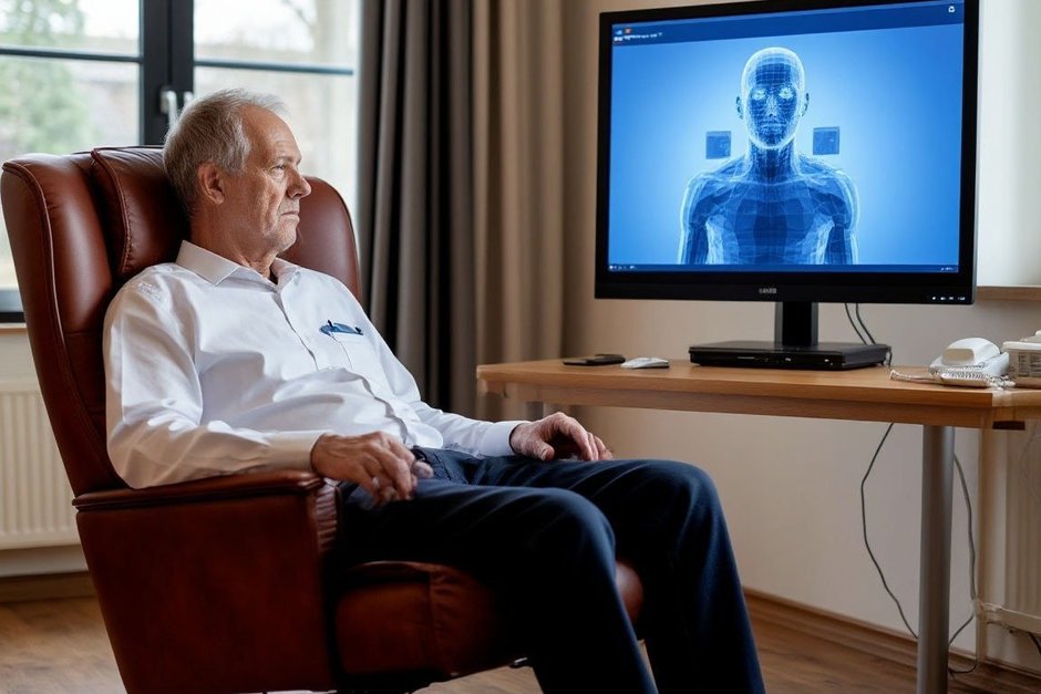
The system translated the recognized speech into text, and the recognized gestures made the digital avatar move. Thus, the participant could, for example, “say” something and simultaneously nod — and words appeared on the screen, and the avatar repeated the movement.
In a conversational experiment one participant achieved an average accuracy of 75% for speech and 85% for gestures. For another the median accuracy of recognition of both speech and gestures was 100%. At the same time the system rarely took the absence of action for an attempt to say or do something.
However, it is still far from the practical application of the technology. Currently the system works with a limited set of pre-given phrases and gestures, and its use requires a surgically installed implant. Besides, the study was small — a full-fledged check of the technology will require trials with a larger number of participants and a much broader vocabulary.
Nevertheless, the experiment shows an important principle: a single implant may be enough to simultaneously read different intentions of a person and turn them into a more natural form of communication — not only into printed words, but also into a digital “body language” that is so far not colored by intonation.